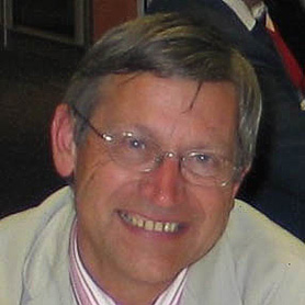
Dr. Paul Lecoq
Senior Physicist at CERN, Geneva, Switzerland and Technical Director of European Center for Research in Medical Imaging in Marseilles
Lectures
Development Of New Scintillating Crystals For High Energy Physics, Medical Imaging And Other Applications
Scintillating crystals have been for a long time developed as a basic component in particle detectors with a strong spin-off in other fields like medical imaging, homeland security and oil well lodging. A typical example is BGO, which has become the main component of PET scanners since the large effort made by the L3 experiment at CERN to develop low cost production methods for this crystal.
Systematic studies on basic mechanisms in inorganic scintillators was initiated by the Crystal Clear Collaboration at CERN 20 years ago, in the frame of a large R&D program to develop the detector technologies for the new CERN proton-proton collider, the LHC. The very special requirements of the scintillating crystals for the Electromagnetic Calorimeter at the CERN Large Hadron Collider CMS experiment have been the subject of intensive research and development. At the start of these studies it was by no means clear that the very high purity of raw material, nor the special and harsh requirements regarding the radiation hardness of these crystals could be met at all. None of the most experienced manufacturers in the field was at that time anywhere close to being able to deliver the quality of crystals needed. This large multidisciplinary effort has contributed not to a small amount, to the development of new materials and new production methods for a new generation of detectors with increased resolution and sensitivity. Some examples will be given in this talk for different application areas.
Spin-Off From Particle Detectors In The Field Of Medicine And Biology
Since the discovery of X-Rays by Roentgen in 1895 physicists have played a major role in the development of medical imaging instrumentation. More recently the technological developments in several areas of applied physics, the new generation of particle physics detectors and the development of an information based society all combine to enhance the performance of presently available imaging devices.
This talk will explain the critical parameters of modern medical imaging in the context of the spectacular development of in-vivo molecular imaging, which will soon allow to bridge post-genomics research activities with new diagnostics and therapeutic strategies for major diseases. In particular the molecular profiling of tumours and gene expression open the way to tailored therapies and therapeutic monitoring of major diseases like cancer, degenerative and genetic disorders. Moreover, the repeatability of non-invasive approaches allows an evaluation of drug targeting and pharmacokinetics studies on small animals, as well as a precise screening and treatment follow-up of patients. The technical requirements on imaging devices are very challenging but are rather similar in many respects to the ones of modern particle detectors on high luminosity accelerators. Examples will be given of active technology transfer areas from High Energy Physics detectors, which can significantly improve the performance of future medical imaging devices.
Special emphasis will be put on the need for a globalisation of technology research and development as modern instrumentation in a vast range of applications has similar requirements and spin-off should be more and more understood as cross-fertilization between different disciplines.
Metamaterials For Novel X Or Gamma Ray Detector Designs
In the majority of X and gamma ray conversion detector heads there is generally a trade-off between the spatial and the energy resolution, as a good spatial resolution requires a high segmentation whereas a good energy resolution is obtained in a large enough detector volume to contain all the cascade interactions generated by the incoming particle. The quest for better spatial resolution in all three dimensions for the majority of applications (High-energy physics and particle detectors, Spectrometry of low energy gamma- quanta, Medical imaging, Homeland security, Space applications) may lead to a huge increase of the number of readout channels, with all the associated problems of connectivity, detector integration and heat dissipation.
This talk will explore the potential of recent progress in the field of crystallogenesis, quantum dots and photonics crystals to develop a new concept of X- and Gamma-ray detector based on metamaterials to simultaneously record with high precision the maximum of information of the cascade conversion process such as its direction, the spatial distribution of the energy deposition and its composition in terms of electromagnetic, charged and neutral hadron contents (for high energy).
Molecular Imaging Challenges With PET and SPECT Techniques
The future trends in molecular imaging and associated challenges for in-vivo functional imaging will be illustrated on the basis of a few examples, such as atherosclerosis vulnerable plaques imaging or stem cells tracking. A set of parameters will be derived to define the specifications of a new generation of in-vivo imaging devices in terms of sensitivity, spatial resolution and signal to noise ratio. The limitations of strategies used in present PET and SPECT scanners will be discussed and new approaches will be proposed taking advantage of recent progress on materials, photodetectors and readout electronics. A special focus will be put on metamaterials, as a new approach to bring more functionality to detection devices. It will be shown that the route is now open towards a fully digital detector head with very high photon counting capability over a large energy range, excellent timing precision and possibility of imaging the energy deposition process.
How to Improve Timing Resolution in Scintillators
The renewal of interest for Time of Flight Positron Emission Tomography (TOF PET), as well as the necessity to precisely tag events in High Energy Physics (HEP) experiments at future colliders, where high luminosity is achieved through high density trains of bunches are pushing for an optimization of all factors affecting the time resolution of the whole acquisition chain: crystal, photodetector, electronics.
The time resolution of a scintillator-based detection system is determined by the rate of photoelectrons at the detection threshold, which depends on the time distribution of photons being converted in the photo-detector.
The possibility to achieve time resolution of about 100ps requires an optimization of the light production in the scintillator, the light transport and its transfer from the scintillator to the photodetector. In order to maximize the light yield, and in particular the density of photons in the first nanosecond, while minimizing the rise time and decay time a particular attention must be given to the energy transfer mechanisms to the activator as well as to the energy transition type at the activator ion.
A particular emphasis will be put on the light transport within the crystal and the transfer to the photo-detector. Light being produced isotropically in the scintillator the detector geometry must be optimized to decrease the optical path-length to the photodetector. Moreover light bouncing within the scintillator must be reduced as much as possible. It concerns typically about 70% of the photons generated in currently used scintillators. It will be shown how photonics crystals specifically designed to couple light propagation modes inside and outside the crystal at the limit of the total reflection angle can significantly improve this situation and impact on the time resolution. Examples of production and deposition of photonics crystals on LYSO crystals will be shown as well a first results on light extraction improvement.
Goals and Achievements of the EndoTOFPET-US FP7 Project
EndoTOFPET-US is an approved European FP7 multidisciplinary project involving an international collaboration of 6 academic institutions (CERN, DESY, Delft Technical University, Lisbon LIP laboratory, University of Heidelberg, University Milano Biccoca), 3 university hospitals (Marseilles Timone, Lausanne CHUV, Münich Technical University hospital) and 3 companies (Fibercryst, KLOE, Surgiceye).
The main clinical objective is the development of new biomarkers for the prostate and the pancreatic cancer and more generally image-guided diagnosis and minimally invasive surgery.
In the frame of this project it is proposed to design and build one prototype of a bi-modal PET-US (Positron Emission Tomography and Ultrasound) endoscopic probe combining in a miniaturized system a fully digital, 200ps time resolution Time of Flight PET detector head (TOF-PET) coupled to a commercial ultrasound (US) assisted biopsy endoscope and to launch a pilot clinical study focusing on pancreatic cancer, after a first step of preclinical feasibility tests on pigs. As an example of novel development of biomarkers, promising antibodies already developed for pancreatic cancer will be pushed towards clinical application.
In order to achieve this very ambitious goal this project will implement a number of novel technologies, among which a new generation of fully digital SiPM photodetectors with single optical photon counting capability, a very compact diffractive optics coupling system between the crystal and the photodetector to compensate for the reduced fill-factor of the later, a low noise time over threshold front end electronics based on the NINO chip developed at CERN for the LHC ALICE experiment and an elaborate tracking system to reconstruct in real time the six coordinates of the internal endoscopic probe and the external plate of the PET detection system.
First performance results of these different components will be presented, including an impressive coincidence time resolution of 155 to 210ps FWHM obtained with crystals of realistic dimensions for the PET (for a length ranging between 5mm and 20mm respectively) using the NINO electronics and a commercial, not yet digital SIPM photodetector.
About

Paul Lecoq has received his diploma as engineer in physics instrumentation at the Ecole Polytechnique de Grenoble in 1972, under the leadership of Nobel Laureate Louis Néel. After two years of work at the Nuclear Physics laboratory of the University of Montreal, Canada, he got his PhD in Nuclear Physics in 1974. Since then he has been working at CERN in 5 major international experiments on particle physics, two of them led by Nobel Laureates Samuel Ting and Carlo Rubbia. His action on detector instrumentation, and particularly on heavy inorganic scintillator materials has received a strong support from Georges Charpak. Member of a number of advisory committees and of international Societies he is since 2002 the promoter of the European Center for Research in Medical Imaging (Cerimed) presently being installed in Marseilles. He is an elected member of the European Academy of Sciences (2008).